mmu-miR-181c at P60 |
|
| Annotation Summary | |
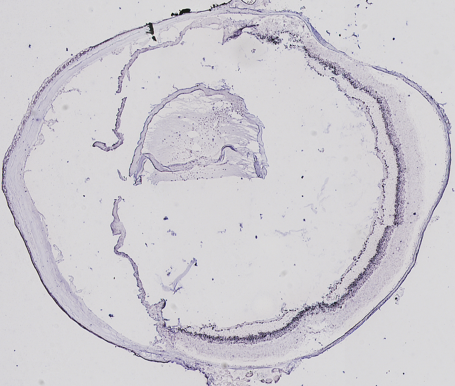
| Anatomical Structures | "GCL, INL" |
| Go with the mouse on the picture to see area zoom | |
|
|
The expression patterns obtained were annotated based on the overall signal distribution and on the anatomical structures and/or cell-types that stained positive for miRNA expression. The terms and abbreviations used for the annotation are described briefly below.
nd: ‘not detected’; term applied in the absence of a visible signal.
reg/1-2-3: ‘regional’; term applied whenever the signal was restricted to specific cells or structures.
ubi/1-2-3: ‘ubiquitous’; term applied when a uniform signal was visible across the entire section.
uwp/1-2-3: ‘ubiquitous with pattern’; term applied for head sections at E16.5 and P0 in cases where a signal was detected in most cells with some structures showing stronger staining.
…/1-2-3: numbers represent a scale of approximate signal intensity. (Please note that RNA in situ hybridization does not provide absolute quantitative data.)
BR = Brain
CB = Ciliary body
GCL = Ganglion Cell Layer
INBL = Inner Neuroblastic Layer
INL = Inner Nuclear Layer
IR = Iris
OLF = Olfactory epithelium
ONBL = Outer Neuroblastic Layer
ONL = Outer Nuclear Layer
PHOT = Photoreceptors
RPE = Retinal Pigment Epithelium
mmu-miR-181c at P60 |
|
| Annotation Summary | |
| Anatomical Structures | "GCL, INL" |
| Go with the mouse on the picture to see area zoom | |
|
|
|